Cheek brightfield Cheek biologycorner cells Human cheek cell dna extraction
Human Cheek Cells Under the Microscope | Haematoxylin | Cell Membrane
Cheek cell human temporary stained cells mounts prepare epithelial lab results layer work discussion study Cheek cell lab – hailey's blog To prepare stained temporary mounts of human cheek cell
Cheek dna extraction chromosomes mugeek vidalondon genetic
Cheek cell bacteria cells human nucleus membrane using bacterial single been prokaryotic solved determineCheek stained microscope My opera is now closedCells cheek microscope human under cell do animal membrane epithelium.
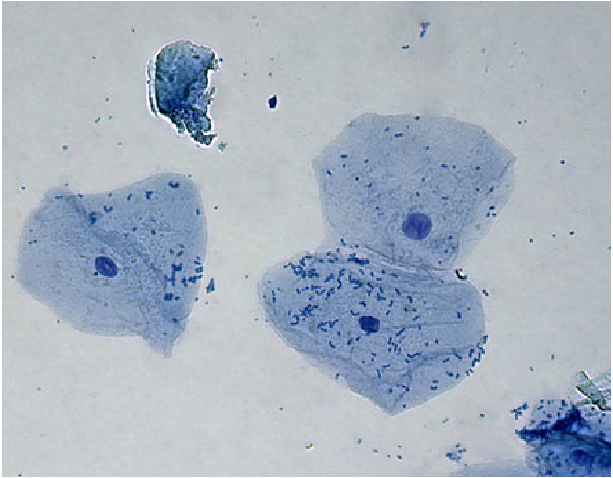
Cheek cell image using brightfield and darkfield microscopy. (aCheek cells under the microscope Cheek onion cell vs cells comparing contrastingDna cells cheek isolation human.

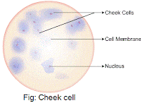
Label the following parts of human cheek cell
Cheek cell human draw labelling correctDraw the diagram of cheek cells and label the parts. Cheek cell human label parts brainly following answerIsolation of dna from human cheek cells.
Cheek cell image using brightfield and darkfield microscopy. (aMicroscopy darkfield brightfield cheek Cells cheek microscope answers 400x staining aim observations eukaryotes schoolworkhelper methylene biology microscopes activities zelula quatr nucleus rosenCheek microscope under cells.
How would you take the sample to prepare temporary stained mount of
Diagram of. cheek cellHuman cheek cells under the microscope Draw the human cheek cell with correct labellingCells cheek human microscope 40x scp cell under 1809 stained 400x magnification blue swab total microscopic stain unstained thf biological.
Solved using this table from the size estimation module,Cheek cell under 40x 400x magnification cells lab nucleus nose piece Human cheek cell ( class : 8 lesson no : 8 )Plant & animal cells staining lab answers.


Human Cheek Cells Under the Microscope | Haematoxylin | Cell Membrane

Cheek cell image using brightfield and darkfield microscopy. (a

Isolation of DNA from Human Cheek Cells - YouTube

PPT - Onion vs. Cheek Cell PowerPoint Presentation, free download - ID

label the following parts of human cheek cell - Brainly.in

How would you take the sample to prepare temporary stained mount of
To prepare stained temporary mounts of human cheek cell - Lab Work

cell-cheek-03 | Cheek Cells | biologycorner | Flickr

Cheek cells under the Microscope - YouTube